TCR Tetramers
MBL has developed and manufactured various major histocompatibility complex (MHC) Tetramer products as tools for detecting antigen-specific T cells. These products have been used to monitor T cell immune responses in research studies and clinical trials.
MBL has efficiently manufactured a TCR Tetramer reagent, which is recognized by MHC, and has successfully commercialized it. Moreover, our cancer antigen-specific (survivin-2B, PBF) TCR Tetramer reagents* have also been assessed for their staining properties by flow cytometry.
*These products are licensed under JP patent No. P2017-169566A.

What is a TCR Tetramer?
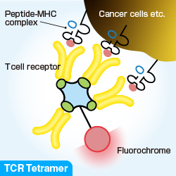
A TCR Tetramer is a reagent prepared by tetramerizing biotinylated antigen-specific TCRs with the help of phycobiliprotein-labeled streptavidin.
Product Highlights
| Product Code | PRODUCT NAME |
|---|---|
| TS-TCR01-1 | Human TCR Tetramer-PE (HLA-A*24:02 survivin-2B80-88-AYACNTSTL) |
| TS-TCR02-1 | Human TCR Tetramer-PE (HLA-A*24:02 PBF145-153-AYRPVSRNI) |
Preparation of TCR Tetramers
The manufacturing process is outlined in the figure below. Purified recombinant TCR α and β chains are refolded for several days to obtain soluble monomeric TCRs (monomers). The refolded monomer is conjugated with a single biotin molecule as the expressed recombinant β chain is already biotinylated at the C-terminal end. The biotinylated monomers were purified and subsequently linked by adding fluorochrome-labeled streptavidin.

Comparison between MHC and TCR Tetramers
| MHC TETRAMER | TCR TETRAMER | |
|---|---|---|
| Structure | MHC/peptide | TCR (α chain/β chain) |
| Recognition | Specific TCR | Specific MHC/peptide complex |
| Detection | Antigen-specific T cells | Antigen-specific tumors and antigen presenting cells |
| Application | ・Quantification ・Monitoring of antigen-specific T cells |
・Quantification ・Evaluation of the efficacy of cancer immunotherapies ・Quality control of cancer vaccine |
Staining using TCR Tetramers
Example #1: Cell Staining (Survivin-2B TCR Tetramer)
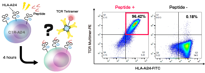
C1R cells (HLA-A-deficient) that stably expressed HLA-A*24:02 (called C1R-A24), were prepared; these cells were pulsed with survivin-2B peptide (AYACNTSTL) for 4 hours. Thereafter, the cells were stained with PE-labeled survivin-2B TCR Tetramer and Anti-HLA-A24 (Human) mAb-FITC (Code No. K0209-4) and further analyzed by flow cytometry. 96.42% of HLA-A24+/Tetramer+ cells were detected while staining peptide-pulsed C1R-A24 cells.

Numbers in the upper right quandrants represent the percentages of TCR Tetramer+ cells relative to the total HLA-A24+ cells
Example #2: Staining of MHC Tetramer-conjugated beads (Survivin-2B TCR Tetramer)
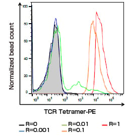
Streptavidin-coated beads were loaded with biotinylated peptide/HLA complex (HLA-A*24:02 presenting survivin-2B peptide, AYACNTSTL) at dilution ratios ranging from 1 to 10−3. This mimics the difference in the pMHC expression in cells.

The following data reveal the staining of the beads using Survivin-2B TCR Tetramer. A representative histogram indicates TCR Tetramer-positive populations in the normalized bead count. The noncoated beads were used as a negative control (R=0).
Reactivity of TCR Tetramer depends on the number of peptide/HLA complexes coated on the beads.


